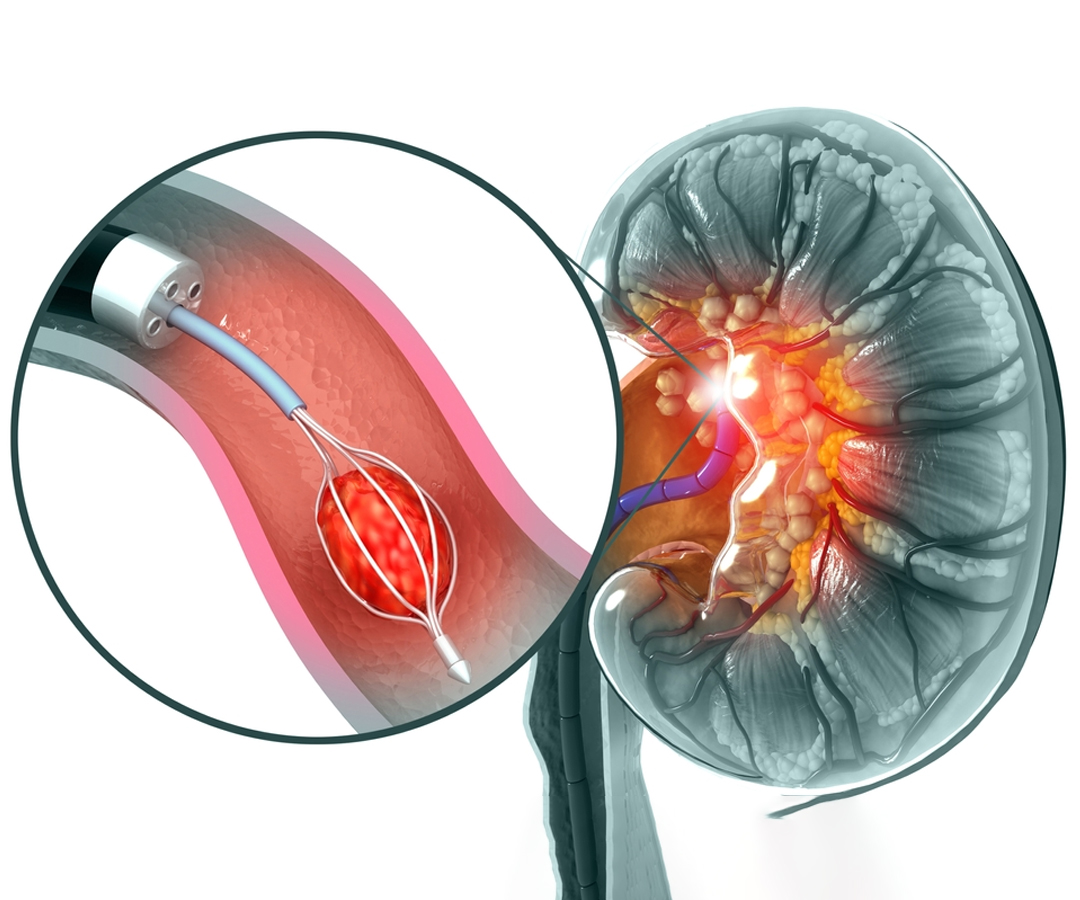
How Can We Suspect Stone Disease?
Kidney stones typically present with severe pain, often starting in the flank region and
radiating down to the groin or genital area. This pain can be intense, and the patient
may feel nauseous or even vomit. Despite tossing and turning in bed, the pain remains
unrelieved. Other symptoms may include a burning sensation while urinating, frequent
urination with small amounts of urine, and occasionally blood in the urine. A fever with
chills is a serious sign of infection and should not be ignored.
In some cases, kidney stones may silently damage the kidney, causing significant
complications. A stone may block the drainage from the kidney, leading to hydronephrosis
(swelling of the kidney). In extreme cases, this can result in complete loss of kidney
function.
How Is the Diagnosis of Stone Disease Made?
When there is a suspicious history or non-specific abdominal pain, diagnostic tests are
needed to confirm the presence of kidney stones. Stones larger than a few millimeters
can often be detected using ultrasound and X-rays. However, these methods have certain
limitations and may miss smaller stones. A plain CT scan of the abdomen is highly
accurate, with up to 98% sensitivity in detecting even the smallest stones, including
those that may not be visible on X-rays. For proper evaluation, patients may require
specialized radiological tests such as IVP (Intravenous Pyelogram) or CT-IVP before
considering any surgical intervention.
When Should Kidney Stones Be Treated?
While kidney stones are common, many smaller stones or crystals pass through the urinary
system without causing major problems. However, as the size of the stone increases or as
it moves further from the bladder, the likelihood of spontaneous clearance decreases.
Small renal stones located on the periphery of the kidney can often be safely monitored
with periodic ultrasound examinations. However, some stones require surgical removal if
they cannot be managed with observation or medication.
Indications for Treatment
- Small stones obstructing urine flow from the kidneys
- Large (staghorn) stones in the kidneys
- Stones in the upper ureter causing pain
- Ureteral stones greater than 5 mm causing pain
- Bladder stones larger than 1 cm
- Stones in a single kidney (whether congenital or following the removal of the
opposite kidney)
- Infected stones
- Recurrent urinary tract infections (UTIs) due to stones
- Stones in individuals working as pilots or in the military, where frequent movement
or strain is involved

